Phone Appointment
[smartblock id=45]WhatsApp Appointment
[smartblock id=46]Most women with breast cancer have some type of surgery as part of their treatment. Your doctor may recommend an appropriate operation based on the size and stage of your cancer, your other treatment options, and your goals and preferences.
Surgery Options
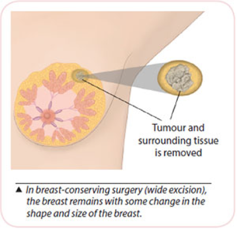
Breast Conserving Surgery (BCS) a.k.a. Wide Excision / Lumpectomy
- Your surgeon will remove only the breast cancer and a rim of normal surrounding breast tissue.
- Your breast will remain. A scar and some changes in shape and size are expected.
- You can go home the next day.
- You may require a 2nd operation if cancer cells are noted at the edge of the removed portion. This occurs in 10-15% of patients.
- You will need to have radiation therapy to the breast (Mon to Fri) for 4- 6 weeks. This helps to destroy any remaining cancer cells.
Oncoplastic Breast Conserving Surgery Mammoplasty (Breast Lift / Breast Reduction)
- All the points for BCS mentioned above are applicable.
- In addition, you will benefit from re-shaping of the breast after cancer removal.
- This is performed to reduce breast deformity and improve the final appearance of the breast after cancer removal.
- Because your cancer is large, the final breast volume may be smaller. Surgery to the opposite breast for better symmetry may be performed at the same time, or at a later date.
- Speak to your surgeon for specific details of this surgery.
Volume Replacement with a Perforator Flap
- All the points for BCS mentioned above are applicable.
- You are suitable for volume replacement with breast conserving surgery.
- Fatty tissue next to your breast will be used to fill the space in the breast that results from cancer removal.
- This maintains breast volume and contour, reducing breast deformity.
- Speak to your surgeon for specific details on this surgery.
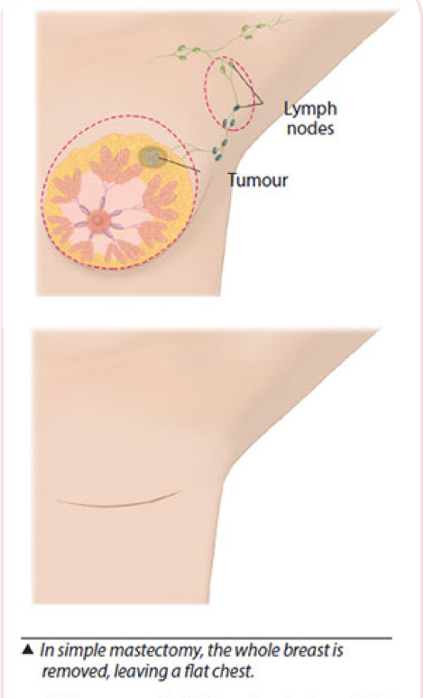
Simple Mastectomy
- The entire breast, including the nipple-areola complex. After surgery, a neat and flat chest wound is expected.
- A soft tubing known as a drain is placed during surgery with the accompanying bottle to remove blood and fluid accumulated at the operated site.
- The drain is removed when the drainage is less than 30ml/24h after 1 to 2 weeks. You will be taught how to care for the drain.
- You can go home the next day.
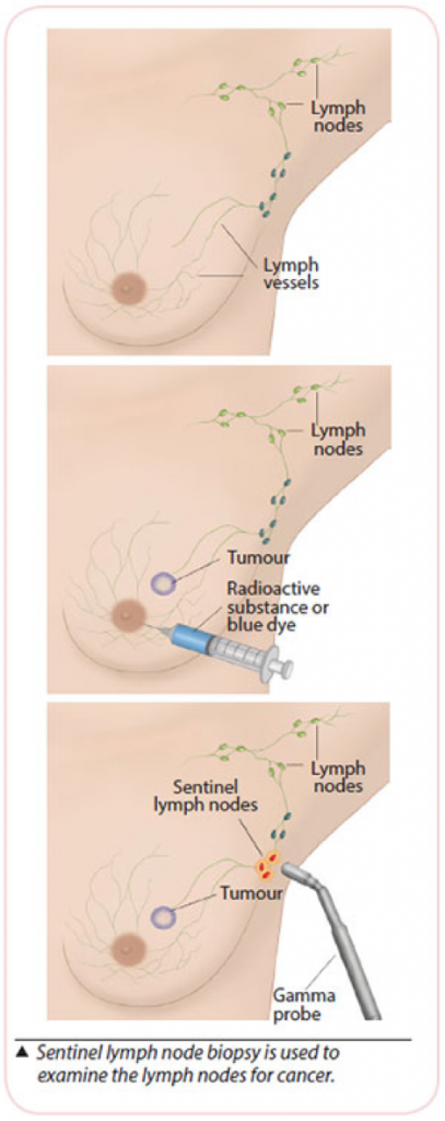
Sentinel Lymph Node Biopsy (SLNB)
- You have early stage breast cancer, and the lymph nodes in your underarm do not appear to have cancer.
- The first few lymph nodes (sentinel lymph nodes, SLN) in your underarm where the lymphatic vessels from the breast drain to, will be removed and examined during surgery under the microscope (frozen section).
- This is done under GA (asleep) and it is to determine if cancer have spread to these SLN.
- A blue dye or a radioactive substance is injected around the cancer site or at the nipple prior to surgery to locate the SLN. The radioactive substance will be injected before the operation in the Department of Nuclear Medicine.
- The blue dye will be injected during the operation.
- If cancer is detected in the SLN, lymph nodes in the axilla are removed. If no cancer is detected in the SLN, no further surgery is needed.
- The final histology will be reviewed about 1 week after surgery.
- In 2-3% of cases, the final assessment of the SLN may be different from the initial frozen section result and a second operation may be required.
- If the dye or radioactive substance are not able to identify the SLN and we will have to proceed to perform an axillary clearance.
Axillary Clearance
- Involves the removal of all lymph nodes from the underarm.
- This is needed because the lymph nodes are found to have cancer cells.
- Side Effects:
- Shoulder stiffness.
- Swelling of the arm (lymphedema) 15-20%.
- Numbness of the inner part of your upper arm.
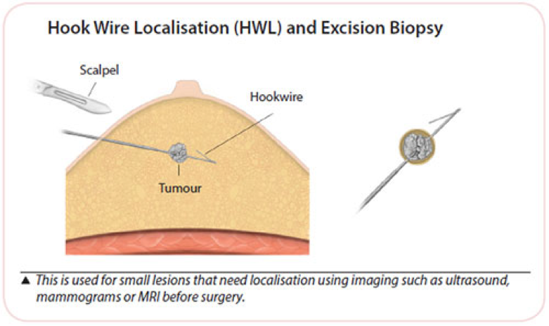
X-ray Guided or Ultrasound Guided Hookwire Localisation for Surgery
- This procedure is performed under local anaesthesia, before you go for surgery.
- Mammogram or ultrasound guidance is used to place a fine wire within the breast, within or in close proximity to the lesion of interest.
- This wire marks the lesion to be removed.
- The wire and lesion of interest will be removed during the surgery.