Phone Appointment
[smartblock id=45]WhatsApp Appointment
[smartblock id=46]A lump in your breast may not necessarily mean cancer. To determine, whether a lump is benign or malignant, biopsies are sometimes performed to enable the doctor to study the tissue and determine if it is cancerous.
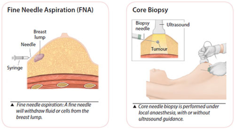
Fine Needle Aspiration (FNA)
A syringe with a very fine needle is used to withdraw fluid or cells from a breast lump. This is a simple procedure and can be uncomfortable but is usually tolerable enough for it to be done in the clinic.
If the lump is just a cyst, withdrawing fluid in this manner will usually make the cyst disappear.
However, if the lump is solid, your doctor may use this procedure to withdraw some cells from it. The cells will then be sent to a laboratory for examination.
Core Needle Biopsy
This is a minimally invasive method that obtains a few tiny strips of tissue from an area of abnormality with a wide bore needle. Local anaesthetic is injected to numb the breast area, followed by a small incision in the skin to allow easy insertion of the needle.
If the abnormality is non- palpable (not detectable by clinical examination) and visible on the ultrasound, ultrasound guidance is used to obtain the tissue. Usually 2 to 6 cores of tissue will be obtained for examination.
A nurse will apply compression to the breast to stop any bleeding. The wound is closed by a steristrip and the dressing applied. Strenuous activity is to be avoided for 2 days after the biopsy.
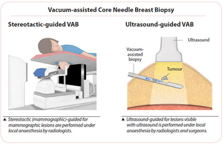
Vacuum-assisted Core Needle Breast Biopsy
Vacuum-assisted biopsy (VAB) devices use a larger bore needle with a vacuum component to obtain tissue samples from non-palpable lesions.
Like the usual core biopsy, this minimally invasive procedure is also performed under local anaesthesia, which is injected to numb the breast area, followed by a small incision in the skin to allow easy insertion of the needle. It is used for lesions seen by mammography (stereotactic-guided biopsy), ultrasound or MRI.
The surgeon or radiologist places the probe into the suspicious area of the breast accurately. A vacuum then draws the tissue into the probe, a cutting device removes the tissue sample and then carries it through the probe into a collection area. More tissue is usually obtained using the VAB than the usual core needle biopsy and the number of strips removed is dependent on the area that needs to be examined.
A small titanium clip (microclip) may be placed at the biopsy site as a location marker for future treatment. This clip is very small (2 mm), is harmless, and will not cause any problems when left inside the breast. An X-ray is taken post-biopsy to ensure proper clip placement.
A nurse will apply compression to the breast to stop any bleeding, the wound is closed by a steristrip and the dressing applied. Strenuous activity is to be avoided for 2 days after the biopsy.
This procedure is minimally invasive as compared to an open surgical biopsy. It is performed as a day surgery procedure. lt has the ability to sample tiny abnormalities called microcalcifications, making early diagnosis of breast cancer possible.
Under local anaesthesia, it takes about 30 to 45 minutes to complete. The procedure is usually not painful but you may experience some discomfort.